CBCT (3D Xray)
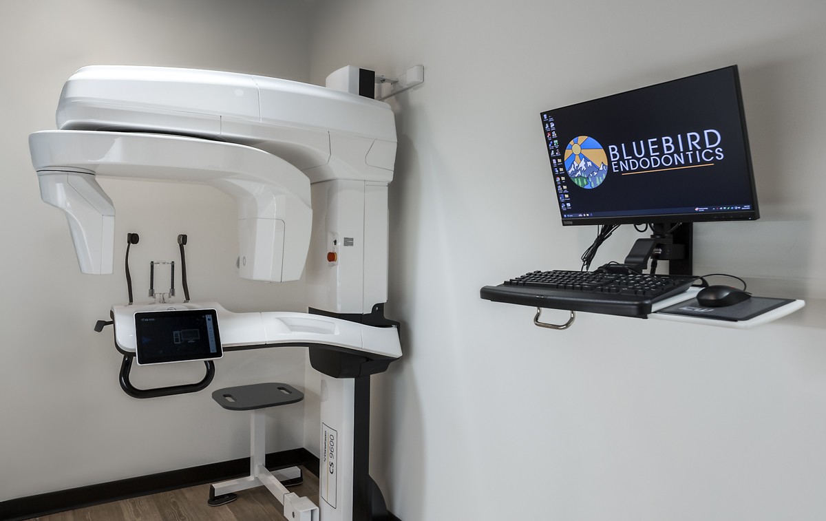
Our office offers the latest in 3-D imaging also known as Cone Beam Computed Tomography (CBCT).
Dr. Heyse uses the 3-D image of your tooth to aid with the following aspects of your care:
- Diagnosis: When a patient presents with complex dental issues or a difficult-to-diagnose problem, a CBCT scan is used to obtain detailed, three-dimensional images of the teeth, roots, surrounding bone, and related structures. These images provide a comprehensive view of the dental anatomy, helping identify the source of any problem accurately.
- Treatment Planning: CBCT scans assist in developing an effective treatment plan. By visualizing the internal structures, Dr. Heyse can identify the number and position of root canals, determine the presence of additional canals, and evaluate the complexity of the tooth's anatomy. This information guides Dr. Heyse in choosing the appropriate techniques and instruments required for a successful root canal treatment.
- Assessing Anatomy: CBCT scans enable the endodontist to evaluate the morphology of root canals and their relationship to adjacent structures. It helps identify the presence of calcified canals, internal resorption, fractures, and other complications that may impact the treatment outcome. This detailed anatomical information allows Dr. Heyse to plan the procedure accordingly and anticipate any challenges that may arise during treatment.
- Localization of Lesions/Infections: In cases where the source of pain or infection is not evident from conventional X-rays or clinical examination, a CBCT scan can assist in localizing lesions such as periapical abscesses, cysts, or other pathologies.
- Evaluation of Treatment Success: After performing root canal treatment, the endodontist may utilize CBCT imaging for post-treatment evaluation. By comparing the pre- and post-treatment scans, they can assess the quality of the treatment, confirm the complete filling of the canals, and verify the resolution of any previously observed pathologies.

Surgical Operating Microscope
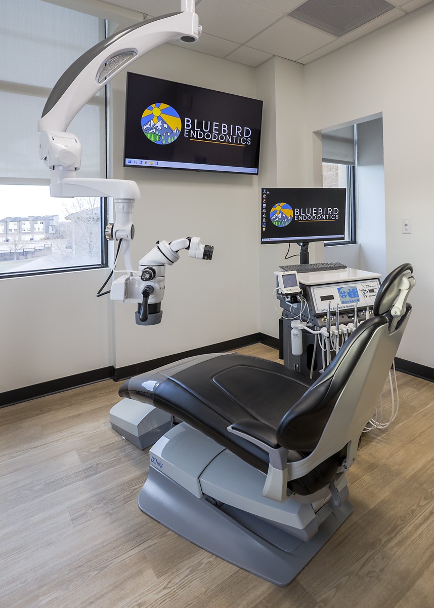
The introduction of the surgical microscope has revolutionized the field of endodontic microsurgery. We have invested in the very best quality surgical microscopes that provide unparalleled magnification and illumination for our surgical procedures.
Dr Heyse uses a microscope for all of his treatments. The advantage of doing root canals through a microscope over dental loupes are numerous. The dental microscope provides the following:
- Enhanced Visualization: The microscope provides significantly improved visualization compared to the naked eye or traditional dental loupes. It offers high magnification and illumination, allowing Dr Heyse to see intricate details of the tooth's anatomy, including the root canals, pulp chamber, and tiny structures within them. This enhanced visualization helps in identifying additional canals, calcifications, fractures, and other complexities that may not be visible otherwise.
- Precise Treatment: Root canal treatment requires precision and accuracy. The microscope enables the endodontist to perform procedures with exceptional precision, as they can see the treatment area in great detail. This is particularly important when working in the narrow and curved canals of the tooth. The endodontist can navigate the complex canal anatomy, locate and remove infected tissue, and shape the canals more effectively, leading to improved treatment outcomes.
- Detection of Microscopic Defects: Microscopic fractures or defects in the tooth structure can be challenging to detect without magnification. The microscope helps Dr Heyse identify such subtle defects that may contribute to the patient's symptoms or impact the success of the treatment. By identifying these issues early, we can address them appropriately during the treatment process.
- Greater Accuracy in Treatment Assessment: After completing the root canal procedure, the endodontist uses the microscope for detailed assessment and evaluation. They can inspect the canals and confirm the thorough cleaning and shaping of the roots. The microscope assists in detecting any residual infected tissue or calcifications that may require further treatment. This level of accuracy in assessment contributes to a higher success rate for the root canal treatment.
- Documentation and Patient Education: The microscope allows the endodontist to capture images or videos of the treatment process. These visual records serve as valuable documentation for future reference or for communication with other dental professionals if necessary. Moreover, the endodontist can share these images with patients, helping them understand the nature of their dental condition, the need for treatment, and the steps involved. This visual aid improves patient education and enables them to make informed decisions about their oral health.

TDO Software
We use TDO Software as it is considered the best endodontic software available. It is used to manage all patient records and information and has comprehensive modules that make our office paperless, a great convenience for our patients and referring dentists. The website integration allows our patients to securely access the site to complete the medical history and consent forms online before their appointment. The software allows our referring dentists to make referral and receive their patients’ reports and imaging through secured HIPAA compliant portal right after the patient is seen. This technology enables us to diagnose and treat our patients more efficiently and to communicate more effectively with both the patient and referring doctor that is secured and HIPAA compliant.
